21 Jan 2025
Current self-supervised learning methods for 3D medical imaging rely on simple pretext formulations and organ- or modality-specific datasets, limiting their generalizability and scalability. We present 3DINO, a cutting-edge SSL method adapted to 3D datasets, and use it to pretrain 3DINO-ViT: a general-purpose medical imaging model, on an exceptionally large, multimodal, and multi-organ dataset of ~100,000 3D medical imaging scans from over 10 organs. We validate 3DINO-ViT using extensive experiments on numerous medical imaging segmentation and classification tasks. Our results demonstrate that 3DINO-ViT generalizes across modalities and organs, including out-of-distribution tasks and datasets, outperforming state-of-the-art methods on the majority of evaluation metrics and labeled dataset sizes. Our 3DINO framework and 3DINO-ViT will be made available to enable research on 3D foundation models or further finetuning for a wide range of medical imaging applications.
30 Jul 2025

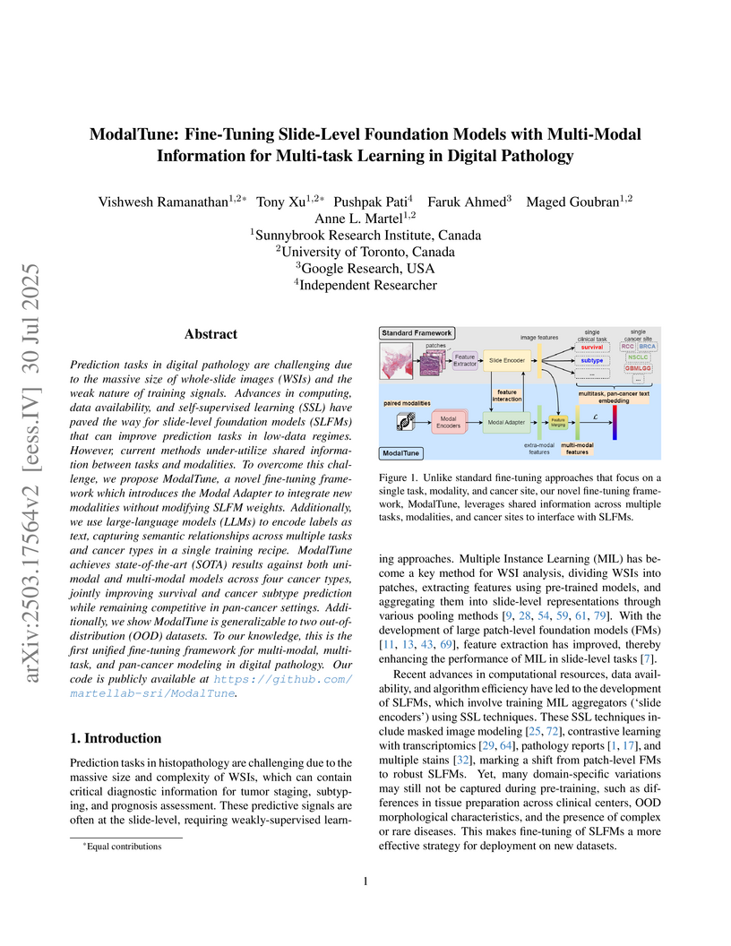
Prediction tasks in digital pathology are challenging due to the massive size of whole-slide images (WSIs) and the weak nature of training signals. Advances in computing, data availability, and self-supervised learning (SSL) have paved the way for slide-level foundation models (SLFMs) that can improve prediction tasks in low-data regimes. However, current methods under-utilize shared information between tasks and modalities. To overcome this challenge, we propose ModalTune, a novel fine-tuning framework which introduces the Modal Adapter to integrate new modalities without modifying SLFM weights. Additionally, we use large-language models (LLMs) to encode labels as text, capturing semantic relationships across multiple tasks and cancer types in a single training recipe. ModalTune achieves state-of-the-art (SOTA) results against both uni-modal and multi-modal models across four cancer types, jointly improving survival and cancer subtype prediction while remaining competitive in pan-cancer settings. Additionally, we show ModalTune is generalizable to two out-of-distribution (OOD) datasets. To our knowledge, this is the first unified fine-tuning framework for multi-modal, multi-task, and pan-cancer modeling in digital pathology.
03 Jun 2023
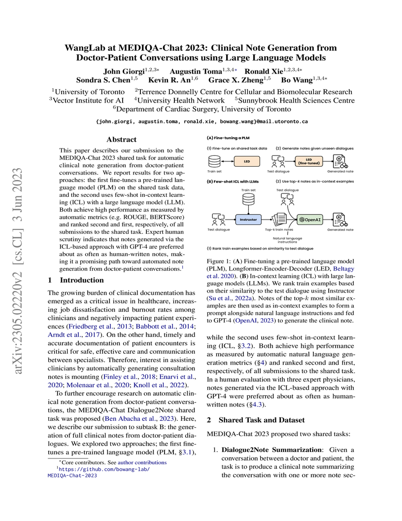
This paper describes our submission to the MEDIQA-Chat 2023 shared task for
automatic clinical note generation from doctor-patient conversations. We report
results for two approaches: the first fine-tunes a pre-trained language model
(PLM) on the shared task data, and the second uses few-shot in-context learning
(ICL) with a large language model (LLM). Both achieve high performance as
measured by automatic metrics (e.g. ROUGE, BERTScore) and ranked second and
first, respectively, of all submissions to the shared task. Expert human
scrutiny indicates that notes generated via the ICL-based approach with GPT-4
are preferred about as often as human-written notes, making it a promising path
toward automated note generation from doctor-patient conversations.
24 Apr 2025
Ordering a minimal subset of lab tests for patients in the intensive care
unit (ICU) can be challenging. Care teams must balance between ensuring the
availability of the right information and reducing the clinical burden and
costs associated with each lab test order. Most in-patient settings experience
frequent over-ordering of lab tests, but are now aiming to reduce this burden
on both hospital resources and the environment. This paper develops a novel
method that combines off-policy learning with privileged information to
identify the optimal set of ICU lab tests to order. Our approach, EXplainable
Off-policy learning with Side Information for ICU blood Test Orders (ExOSITO)
creates an interpretable assistive tool for clinicians to order lab tests by
considering both the observed and predicted future status of each patient. We
pose this problem as a causal bandit trained using offline data and a reward
function derived from clinically-approved rules; we introduce a novel learning
framework that integrates clinical knowledge with observational data to bridge
the gap between the optimal and logging policies. The learned policy function
provides interpretable clinical information and reduces costs without omitting
any vital lab orders, outperforming both a physician's policy and prior
approaches to this practical problem.

30 May 2024
 University of Toronto
 Monash UniversityUniversity of Utah
 University of California, Irvine
 University of California, San DiegoGriffith University
 The Ohio State UniversityThomas Jefferson UniversityWeill Cornell MedicineChina Medical UniversityUniversity of California San FranciscoChiang Mai UniversityMemorial Sloan Kettering Cancer CenterUniversidade Federal de São PauloDuke University School of MedicineYale University School of MedicineVancouver General HospitalSunnybrook Health Sciences CentreEberhard Karls University TübingenMedical College of WisconsinUniversity Hospitals Cleveland Medical CenterMater Dei HospitalAlfred HealthLiverpool HospitalGold Coast University HospitalScripps Clinic Medical GroupSt. Michael’s HospitalUnity Health TorontoChina Medical University HospitalCase Western Reserve University School of MedicineClinical Center University of SarajevoTallaght University HospitalClínica Santa MaríaUniversity Hospital of WürzburgHospital Universitario Ramón y CajalKoç University School of MedicineThe Jackson LaboratoryCHU Mohamed VI Cadi Ayyad UniversityQueens
’ University
The Ohio State UniversityThomas Jefferson UniversityWeill Cornell MedicineChina Medical UniversityUniversity of California San FranciscoChiang Mai UniversityMemorial Sloan Kettering Cancer CenterUniversidade Federal de São PauloDuke University School of MedicineYale University School of MedicineVancouver General HospitalSunnybrook Health Sciences CentreEberhard Karls University TübingenMedical College of WisconsinUniversity Hospitals Cleveland Medical CenterMater Dei HospitalAlfred HealthLiverpool HospitalGold Coast University HospitalScripps Clinic Medical GroupSt. Michael’s HospitalUnity Health TorontoChina Medical University HospitalCase Western Reserve University School of MedicineClinical Center University of SarajevoTallaght University HospitalClínica Santa MaríaUniversity Hospital of WürzburgHospital Universitario Ramón y CajalKoç University School of MedicineThe Jackson LaboratoryCHU Mohamed VI Cadi Ayyad UniversityQueens
’ UniversityThe RSNA Abdominal Traumatic Injury CT (RATIC) dataset is the largest
publicly available collection of adult abdominal CT studies annotated for
traumatic injuries. This dataset includes 4,274 studies from 23 institutions
across 14 countries. The dataset is freely available for non-commercial use via
Kaggle at
this https URL
Created for the RSNA 2023 Abdominal Trauma Detection competition, the dataset
encourages the development of advanced machine learning models for detecting
abdominal injuries on CT scans. The dataset encompasses detection and
classification of traumatic injuries across multiple organs, including the
liver, spleen, kidneys, bowel, and mesentery. Annotations were created by
expert radiologists from the American Society of Emergency Radiology (ASER) and
Society of Abdominal Radiology (SAR). The dataset is annotated at multiple
levels, including the presence of injuries in three solid organs with injury
grading, image-level annotations for active extravasations and bowel injury,
and voxelwise segmentations of each of the potentially injured organs. With the
release of this dataset, we hope to facilitate research and development in
machine learning and abdominal trauma that can lead to improved patient care
and outcomes.
09 Jul 2025

Computational pathology (CoPath) leverages histopathology images to enhance diagnostic precision and reproducibility in clinical pathology. However, publicly available datasets for CoPath that are annotated with extensive histological tissue type (HTT) taxonomies at a granular level remain scarce due to the significant expertise and high annotation costs required. Existing datasets, such as the Atlas of Digital Pathology (ADP), address this by offering diverse HTT annotations generalized to multiple organs, but limit the capability for in-depth studies on specific organ diseases. Building upon this foundation, we introduce ADPv2, a novel dataset focused on gastrointestinal histopathology. Our dataset comprises 20,004 image patches derived from healthy colon biopsy slides, annotated according to a hierarchical taxonomy of 32 distinct HTTs of 3 levels. Furthermore, we train a multilabel representation learning model following a two-stage training procedure on our ADPv2 dataset. We leverage the VMamba architecture and achieving a mean average precision (mAP) of 0.88 in multilabel classification of colon HTTs. Finally, we show that our dataset is capable of an organ-specific in-depth study for potential biomarker discovery by analyzing the model's prediction behavior on tissues affected by different colon diseases, which reveals statistical patterns that confirm the two pathological pathways of colon cancer development. Our dataset is publicly available at this https URL
26 Mar 2025
Lung cancer remains one of the leading causes of cancer-related mortality
worldwide. A crucial challenge for early diagnosis is differentiating uncertain
cases with similar visual characteristics and closely annotation scores. In
clinical practice, radiologists rely on quantitative, hand-crafted Radiomic
features extracted from Computed Tomography (CT) images, while recent research
has primarily focused on deep learning solutions. More recently,
Vision-Language Models (VLMs), particularly Contrastive Language-Image
Pre-Training (CLIP)-based models, have gained attention for their ability to
integrate textual knowledge into lung cancer diagnosis. While CLIP-Lung models
have shown promising results, we identified the following potential
limitations: (a) dependence on radiologists' annotated attributes, which are
inherently subjective and error-prone, (b) use of textual information only
during training, limiting direct applicability at inference, and (c)
Convolutional-based vision encoder with randomly initialized weights, which
disregards prior knowledge. To address these limitations, we introduce
AutoRad-Lung, which couples an autoregressively pre-trained VLM, with prompts
generated from hand-crafted Radiomics. AutoRad-Lung uses the vision encoder of
the Large-Scale Autoregressive Image Model (AIMv2), pre-trained using a
multi-modal autoregressive objective. Given that lung tumors are typically
small, irregularly shaped, and visually similar to healthy tissue, AutoRad-Lung
offers significant advantages over its CLIP-based counterparts by capturing
pixel-level differences. Additionally, we introduce conditional context
optimization, which dynamically generates context-specific prompts based on
input Radiomics, improving cross-modal alignment.
13 Aug 2020
Lung diseases including infections such as Pneumonia, Tuberculosis, and novel
Coronavirus (COVID-19), together with Lung Cancer are significantly widespread
and are, typically, considered life threatening. In particular, lung cancer is
among the most common and deadliest cancers with a low 5-year survival rate.
Timely diagnosis of lung cancer is, therefore, of paramount importance as it
can save countless lives. In this regard, deep learning radiomics solutions
have the promise of extracting the most useful features on their own in an
end-to-end fashion without having access to the annotated boundaries. Among
different deep learning models, Capsule Networks are proposed to overcome
shortcomings of the Convolutional Neural Networks (CNN) such as their inability
to recognize detailed spatial relations. Capsule networks have so far shown
satisfying performance in medical imaging problems. Capitalizing on their
success, in this study, we propose a novel capsule network-based mixture of
experts, referred to as the MIXCAPS. The proposed MIXCAPS architecture takes
advantage of not only the capsule network's capabilities to handle small
datasets, but also automatically splitting dataset through a convolutional
gating network. MIXCAPS enables capsule network experts to specialize on
different subsets of the data. Our results show that MIXCAPS outperforms a
single capsule network and a mixture of CNNs, with an accuracy of 92.88%,
sensitivity of 93.2%, specificity of 92.3% and area under the curve of 0.963.
Our experiments also show that there is a relation between the gate outputs and
a couple of hand-crafted features, illustrating explainable nature of the
proposed MIXCAPS. To further evaluate generalization capabilities of the
proposed MIXCAPS architecture, additional experiments on a brain tumor dataset
are performed showing potentials of MIXCAPS for detection of tumors related to
other organs.
31 Jul 2016

We present and evaluate the capacity of a deep neural network to learn robust
features from EEG to automatically detect seizures. This is a challenging
problem because seizure manifestations on EEG are extremely variable both
inter- and intra-patient. By simultaneously capturing spectral, temporal and
spatial information our recurrent convolutional neural network learns a general
spatially invariant representation of a seizure. The proposed approach exceeds
significantly previous results obtained on cross-patient classifiers both in
terms of sensitivity and false positive rate. Furthermore, our model proves to
be robust to missing channel and variable electrode montage.
28 Mar 2017

Lung cancer is the leading cause for cancer related deaths. As such, there is
an urgent need for a streamlined process that can allow radiologists to provide
diagnosis with greater efficiency and accuracy. A powerful tool to do this is
radiomics: a high-dimension imaging feature set. In this study, we take the
idea of radiomics one step further by introducing the concept of discovery
radiomics for lung cancer prediction using CT imaging data. In this study, we
realize these custom radiomic sequencers as deep convolutional sequencers using
a deep convolutional neural network learning architecture. To illustrate the
prognostic power and effectiveness of the radiomic sequences produced by the
discovered sequencer, we perform cancer prediction between malignant and benign
lesions from 97 patients using the pathologically-proven diagnostic data from
the LIDC-IDRI dataset. Using the clinically provided pathologically-proven data
as ground truth, the proposed framework provided an average accuracy of 77.52%
via 10-fold cross-validation with a sensitivity of 79.06% and specificity of
76.11%, surpassing the state-of-the art method.
28 Feb 2018
Shoulder Physiotherapy Exercise Recognition: Machine Learning the Inertial Signals from a Smartwatch
Shoulder Physiotherapy Exercise Recognition: Machine Learning the Inertial Signals from a Smartwatch
Objective: Participation in a physical therapy program is considered one of
the greatest predictors of successful conservative management of common
shoulder disorders. However, adherence to these protocols is often poor and
typically worse for unsupervised home exercise programs. Currently, there are
limited tools available for objective measurement of adherence in the home
setting. The goal of this study was to develop and evaluate the potential for
performing home shoulder physiotherapy monitoring using a commercial
smartwatch.
Approach: Twenty healthy adult subjects with no prior shoulder disorders
performed seven exercises from an evidence-based rotator cuff physiotherapy
protocol, while 6-axis inertial sensor data was collected from the active
extremity. Within an activity recognition chain (ARC) framework, four
supervised learning algorithms were trained and optimized to classify the
exercises: k-nearest neighbor (k-NN), random forest (RF), support vector
machine classifier (SVC), and a convolutional recurrent neural network (CRNN).
Algorithm performance was evaluated using 5-fold cross-validation stratified
first temporally and then by subject.
Main Results: Categorical classification accuracy was above 94% for all
algorithms on the temporally stratified cross validation, with the best
performance achieved by the CRNN algorithm (99.4%). The subject stratified
cross validation, which evaluated classifier performance on unseen subjects,
yielded lower accuracies scores again with CRNN performing best (88.9%).
Significance: This proof of concept study demonstrates the technical
feasibility of a smartwatch device and supervised machine learning approach to
more easily monitor and assess the at-home adherence of shoulder physiotherapy
exercise protocols.
20 Feb 2019
Recent advancements in signal processing and machine learning coupled with
developments of electronic medical record keeping in hospitals and the
availability of extensive set of medical images through internal/external
communication systems, have resulted in a recent surge of significant interest
in "Radiomics". Radiomics is an emerging and relatively new research field,
which refers to extracting semi-quantitative and/or quantitative features from
medical images with the goal of developing predictive and/or prognostic models,
and is expected to become a critical component for integration of image-derived
information for personalized treatment in the near future. The conventional
Radiomics workflow is typically based on extracting pre-designed features (also
referred to as hand-crafted or engineered features) from a segmented region of
interest. Nevertheless, recent advancements in deep learning have caused trends
towards deep learning-based Radiomics (also referred to as discovery
Radiomics). Considering the advantages of these two approaches, there are also
hybrid solutions developed to exploit the potentials of multiple data sources.
Considering the variety of approaches to Radiomics, further improvements
require a comprehensive and integrated sketch, which is the goal of this
article. This manuscript provides a unique interdisciplinary perspective on
Radiomics by discussing state-of-the-art signal processing solutions in the
context of Radiomics.
10 Jul 2019
As an analytic pipeline for quantitative imaging feature extraction and
analysis, radiomics has grown rapidly in the past a few years. Recent studies
in radiomics aim to investigate the relationship between tumors imaging
features and clinical outcomes. Open source radiomics feature banks enable the
extraction and analysis of thousands of predefined features. On the other hand,
recent advances in deep learning have shown significant potential in the
quantitative medical imaging field, raising the research question of whether
predefined radiomics features have predictive information in addition to deep
learning features. In this study, we propose a feature fusion method and
investigate whether a combined feature bank of deep learning and predefined
radiomics features can improve the prognostics performance. CT images from
resectable Pancreatic Adenocarcinoma (PDAC) patients were used to compare the
prognosis performance of common feature reduction and fusion methods and the
proposed risk-score based feature fusion method for overall survival. It was
shown that the proposed feature fusion method significantly improves the
prognosis performance for overall survival in resectable PDAC cohorts,
elevating the area under ROC curve by 51% compared to predefined radiomics
features alone, by 16% compared to deep learning features alone, and by 32%
compared to existing feature fusion and reduction methods for a combination of
deep learning and predefined radiomics features.
16 Apr 2020
COVID-CAPS: A Capsule Network-based Framework for Identification of COVID-19 cases from X-ray Images
COVID-CAPS: A Capsule Network-based Framework for Identification of COVID-19 cases from X-ray Images
Novel Coronavirus disease (COVID-19) has abruptly and undoubtedly changed the
world as we know it at the end of the 2nd decade of the 21st century. COVID-19
is extremely contagious and quickly spreading globally making its early
diagnosis of paramount importance. Early diagnosis of COVID-19 enables health
care professionals and government authorities to break the chain of transition
and flatten the epidemic curve. The common type of COVID-19 diagnosis test,
however, requires specific equipment and has relatively low sensitivity.
Computed tomography (CT) scans and X-ray images, on the other hand, reveal
specific manifestations associated with this disease. Overlap with other lung
infections makes human-centered diagnosis of COVID-19 challenging.
Consequently, there has been an urgent surge of interest to develop Deep Neural
Network (DNN)-based diagnosis solutions, mainly based on Convolutional Neural
Networks (CNNs), to facilitate identification of positive COVID-19 cases. CNNs,
however, are prone to lose spatial information between image instances and
require large datasets. The paper presents an alternative modeling framework
based on Capsule Networks, referred to as the COVID-CAPS, being capable of
handling small datasets, which is of significant importance due to sudden and
rapid emergence of COVID-19. Our results based on a dataset of X-ray images
show that COVID-CAPS has advantage over previous CNN-based models. COVID-CAPS
achieved an Accuracy of 95.7%, Sensitivity of 90%, Specificity of 95.8%, and
Area Under the Curve (AUC) of 0.97, while having far less number of trainable
parameters in comparison to its counterparts. To further improve diagnosis
capabilities of the COVID-CAPS, pre-training based on a new dataset constructed
from an external dataset of X-ray images. Pre-training with a dataset of
similar nature further improved accuracy to 98.3% and specificity to 98.6%.
30 Oct 2020

The newly discovered Corona virus Disease 2019 (COVID-19) has been globally
spreading and causing hundreds of thousands of deaths around the world as of
its first emergence in late 2019. Computed tomography (CT) scans have shown
distinctive features and higher sensitivity compared to other diagnostic tests,
in particular the current gold standard, i.e., the Reverse Transcription
Polymerase Chain Reaction (RT-PCR) test. Current deep learning-based algorithms
are mainly developed based on Convolutional Neural Networks (CNNs) to identify
COVID-19 pneumonia cases. CNNs, however, require extensive data augmentation
and large datasets to identify detailed spatial relations between image
instances. Furthermore, existing algorithms utilizing CT scans, either extend
slice-level predictions to patient-level ones using a simple thresholding
mechanism or rely on a sophisticated infection segmentation to identify the
disease. In this paper, we propose a two-stage fully-automated CT-based
framework for identification of COVID-19 positive cases referred to as the
"COVID-FACT". COVID-FACT utilizes Capsule Networks, as its main building blocks
and is, therefore, capable of capturing spatial information. In particular, to
make the proposed COVID-FACT independent from sophisticated segmentation of the
area of infection, slices demonstrating infection are detected at the first
stage and the second stage is responsible for classifying patients into COVID
and non-COVID cases. COVID-FACT detects slices with infection, and identifies
positive COVID-19 cases using an in-house CT scan dataset, containing COVID-19,
community acquired pneumonia, and normal cases. Based on our experiments,
COVID-FACT achieves an accuracy of 90.82%, a sensitivity of 94.55%, a
specificity of 86.04%, and an Area Under the Curve (AUC) of 0.98, while
depending on far less supervision and annotation, in comparison to its
counterparts.
30 Oct 2020
The global outbreak of the novel corona virus (COVID-19) disease has
drastically impacted the world and led to one of the most challenging crisis
across the globe since World War II. The early diagnosis and isolation of
COVID-19 positive cases are considered as crucial steps towards preventing the
spread of the disease and flattening the epidemic curve. Chest Computed
Tomography (CT) scan is a highly sensitive, rapid, and accurate diagnostic
technique that can complement Reverse Transcription Polymerase Chain Reaction
(RT-PCR) test. Recently, deep learning-based models, mostly based on
Convolutional Neural Networks (CNN), have shown promising diagnostic results.
CNNs, however, are incapable of capturing spatial relations between image
instances and require large datasets. Capsule Networks, on the other hand, can
capture spatial relations, require smaller datasets, and have considerably
fewer parameters. In this paper, a Capsule network framework, referred to as
the "CT-CAPS", is presented to automatically extract distinctive features of
chest CT scans. These features, which are extracted from the layer before the
final capsule layer, are then leveraged to differentiate COVID-19 from
Non-COVID cases. The experiments on our in-house dataset of 307 patients show
the state-of-the-art performance with the accuracy of 90.8%, sensitivity of
94.5%, and specificity of 86.0%.
13 Jul 2021
We present SurgeonAssist-Net: a lightweight framework making action-and-workflow-driven virtual assistance, for a set of predefined surgical tasks, accessible to commercially available optical see-through head-mounted displays (OST-HMDs). On a widely used benchmark dataset for laparoscopic surgical workflow, our implementation competes with state-of-the-art approaches in prediction accuracy for automated task recognition, and yet requires 7.4x fewer parameters, 10.2x fewer floating point operations per second (FLOPS), is 7.0x faster for inference on a CPU, and is capable of near real-time performance on the Microsoft HoloLens 2 OST-HMD. To achieve this, we make use of an efficient convolutional neural network (CNN) backbone to extract discriminative features from image data, and a low-parameter recurrent neural network (RNN) architecture to learn long-term temporal dependencies. To demonstrate the feasibility of our approach for inference on the HoloLens 2 we created a sample dataset that included video of several surgical tasks recorded from a user-centric point-of-view. After training, we deployed our model and cataloged its performance in an online simulated surgical scenario for the prediction of the current surgical task. The utility of our approach is explored in the discussion of several relevant clinical use-cases. Our code is publicly available at this https URL.
23 Apr 2015
Current image segmentation techniques usually require that the user tune several parameters in order to obtain maximum segmentation accuracy, a computationally inefficient approach, especially when a large number of images must be processed sequentially in daily practice. The use of evolving fuzzy systems for designing a method that automatically adjusts parameters to segment medical images according to the quality expectation of expert users has been proposed recently (Evolving fuzzy image segmentation EFIS). However, EFIS suffers from a few limitations when used in practice mainly due to some fixed parameters. For instance, EFIS depends on auto-detection of the object of interest for feature calculation, a task that is highly application-dependent. This shortcoming limits the applicability of EFIS, which was proposed with the ultimate goal of offering a generic but adjustable segmentation scheme. In this paper, a new version of EFIS is proposed to overcome these limitations. The new EFIS, called self-configuring EFIS (SC-EFIS), uses available training data to self-estimate the parameters that are fixed in EFIS. As well, the proposed SC-EFIS relies on a feature selection process that does not require auto-detection of an ROI. The proposed SC-EFIS was evaluated using the same segmentation algorithms and the same dataset as for EFIS. The results show that SC-EFIS can provide the same results as EFIS but with a higher level of automation.
06 Jan 2023
Purpose: To develop a machine learning-based, 3D dose prediction methodology for Gamma Knife (GK) radiosurgery. The methodology accounts for cases involving targets of any number, size, and shape. Methods: Data from 322 GK treatment plans was modified by isolating and cropping the contoured MRI and clinical dose distributions based on tumor location, then scaling the resulting tumor spaces to a standard size. An accompanying 3D tensor was created for each instance to account for tumor size. The modified dataset for 272 patients was used to train both a generative adversarial network (GAN-GK) and a 3D U-Net model (U-Net-GK). Unmodified data was used to train equivalent baseline models. All models were used to predict the dose distribution of 50 out-of-sample patients. Prediction accuracy was evaluated using gamma, with criteria of 4%/2mm, 3%/3mm, 3%/1mm and 1%/1mm. Prediction quality was assessed using coverage, selectivity, and conformity indices. Results: The predictions resulting from GAN-GK and U-Net-GK were similar to their clinical counterparts, with average gamma (4%/2mm) passing rates of 84.9 and 83.1, respectively. In contrast, the gamma passing rate of baseline models were significantly worse than their respective GK-specific models (p < 0.001) at all criterion levels. The quality of GK-specific predictions was also similar to that of clinical plans. Conclusion: Deep learning models can use GK-specific data modification to predict 3D dose distributions for GKRS plans with a large range in size, shape, or number of targets. Standard deep learning models applied to unmodified GK data generated poorer predictions.
17 Feb 2025
Amyloidosis is a protein misfolding disease caused by the deposition of
large, insoluble aggregates (amyloid fibrils) of protein in a tissue, which has
been associated with various conditions, such as lymphoid disorders,
Alzheimer's disease, diabetes mellitus type 2, chronic inflammatory processes,
and cancers. Amyloid fibrils are commonly diagnosed by qualitative observation
of green birefringence from Congo red stained biopsy tissue samples under
polarized light, a technique that is limited by lack of specificity, dependence
on subjective interpretation, and technical constraints. Studies emphasize the
utility of quantitative polarized light microscopy (PLM) methodology to
diagnose amyloid fibrils in Congo red stained tissues. However, while Congo red
enhances the intrinsic birefringence of amyloid fibrillar structures, there are
significant disadvantages such as the appearance of multiple non-green colors
under polarized light and binding to other structures, which may result in
misdiagnoses with Congo red dye and inconclusive explanations. In this work, we
present an improved PLM methodology for quantitative detection of amyloid
fibrils without requiring Congo red staining. We perform PLM measurements on
four tissues: abdominal subcutaneous tissue biopsy, duodenal biopsy, thyroid
biopsy, and breast biopsy, both with Congo red stain and H\&E stain, and
through Fourier analysis quantify birefringence, birefringent axis orientation,
dichroism, optical activity, and relative amyloid density. These results
emphasize a quantitative analysis for amyloid diagnosis rooted in Fourier
signal harmonics that does not require Congo red dye and paves the way for
rapid, simple, and accurate diagnosis of amyloid fibrils.
There are no more papers matching your filters at the moment.
