28 May 2024
Heidelberg University UCLAUniversity of ZurichGeorge Washington University
 University of California, Irvine
 Stanford University
 University of Michigan
 Cornell University
 University of California, San Diego
 McGill University
 Northwestern UniversityUniversity of Missouri
 University of PennsylvaniaMassachusetts General HospitalUppsala University
 Duke UniversityHelmholtz MunichMayo ClinicWeill Cornell MedicineIran University of Medical SciencesChildren’s National HospitalUniversity Hospital UlmNational Institute of HealthSage BionetworksUniversity of Rochester Medical CenterMercy Catholic Medical CenterKlinikum rechts der Isar, Technical University of MunichThe Children’s Hospital of PhiladelphiaC.M.H. Lahore Medical College & Institute of DentistryRoss University School of MedicineNassau Medical CenterUniversity of LavalIndiana UniveristyHospital Garcia de Orta - Unidade Local de Sa ́ude de Almada Seixal, EPEScripps Clinic Medical GroupHospital da Luz LisboaVA San Diego Medical CenterASST Ovest Milanese - Legnano
UCLAUniversity of ZurichGeorge Washington UniversityUniversity of California, IrvineStanford UniversityUniversity of MichiganCornell UniversityUniversity of California, San DiegoMcGill UniversityNorthwestern UniversityUniversity of MissouriUniversity of PennsylvaniaMassachusetts General HospitalUppsala UniversityDuke UniversityHelmholtz MunichMayo ClinicWeill Cornell MedicineIran University of Medical SciencesChildren’s National HospitalUniversity Hospital UlmNational Institute of HealthSage BionetworksUniversity of Rochester Medical CenterMercy Catholic Medical CenterKlinikum rechts der Isar, Technical University of MunichThe Children’s Hospital of PhiladelphiaC.M.H. Lahore Medical College & Institute of DentistryRoss University School of MedicineNassau Medical CenterUniversity of LavalIndiana UniveristyHospital Garcia de Orta - Unidade Local de Sa ́ude de Almada Seixal, EPEScripps Clinic Medical GroupHospital da Luz LisboaVA San Diego Medical CenterASST Ovest Milanese - LegnanoGliomas are the most common malignant primary brain tumors in adults and one of the deadliest types of cancer. There are many challenges in treatment and monitoring due to the genetic diversity and high intrinsic heterogeneity in appearance, shape, histology, and treatment response. Treatments include surgery, radiation, and systemic therapies, with magnetic resonance imaging (MRI) playing a key role in treatment planning and post-treatment longitudinal assessment. The 2024 Brain Tumor Segmentation (BraTS) challenge on post-treatment glioma MRI will provide a community standard and benchmark for state-of-the-art automated segmentation models based on the largest expert-annotated post-treatment glioma MRI dataset. Challenge competitors will develop automated segmentation models to predict four distinct tumor sub-regions consisting of enhancing tissue (ET), surrounding non-enhancing T2/fluid-attenuated inversion recovery (FLAIR) hyperintensity (SNFH), non-enhancing tumor core (NETC), and resection cavity (RC). Models will be evaluated on separate validation and test datasets using standardized performance metrics utilized across the BraTS 2024 cluster of challenges, including lesion-wise Dice Similarity Coefficient and Hausdorff Distance. Models developed during this challenge will advance the field of automated MRI segmentation and contribute to their integration into clinical practice, ultimately enhancing patient care.
27 Dec 2021
 University of Cambridge
 University of Southern California
 National University of Singapore
 University College London
 University of Oxford
 Georgia Institute of Technology
 University of CopenhagenUniversity of California, San DiegoMcGill University
 Emory UniversityUniversity of Pennsylvania
 Arizona State University
 University of Maryland
 King’s College LondonErasmus MCMayo ClinicLund UniversityBrandeis UniversityBen-Gurion University of the NegevUniversity of Eastern FinlandPortland State UniversityUniversity of California San FranciscoNational Institute on AgingGenentechUniversity of PlymouthBanner Alzheimer’s InstituteUCL Queen Square Institute of NeurologyThe University of Texas Health Science Center at HoustonMedical College of WisconsinGerman Center for Neurodegenerative DiseasesUniversity of GhanaInstituto Tecnol ́ogico y de Estudios Superiores de MonterreyH. Lundbeck A/SVU Medical CentreInstitut du Cerveau et de la Moelle ́epini`ereVasile Lucaciu National CollegeBiomarinIBM Research - Australia
We present the findings of "The Alzheimer's Disease Prediction Of
Longitudinal Evolution" (TADPOLE) Challenge, which compared the performance of
92 algorithms from 33 international teams at predicting the future trajectory
of 219 individuals at risk of Alzheimer's disease. Challenge participants were
required to make a prediction, for each month of a 5-year future time period,
of three key outcomes: clinical diagnosis, Alzheimer's Disease Assessment Scale
Cognitive Subdomain (ADAS-Cog13), and total volume of the ventricles. The
methods used by challenge participants included multivariate linear regression,
machine learning methods such as support vector machines and deep neural
networks, as well as disease progression models. No single submission was best
at predicting all three outcomes. For clinical diagnosis and ventricle volume
prediction, the best algorithms strongly outperform simple baselines in
predictive ability. However, for ADAS-Cog13 no single submitted prediction
method was significantly better than random guesswork. Two ensemble methods
based on taking the mean and median over all predictions, obtained top scores
on almost all tasks. Better than average performance at diagnosis prediction
was generally associated with the additional inclusion of features from
cerebrospinal fluid (CSF) samples and diffusion tensor imaging (DTI). On the
other hand, better performance at ventricle volume prediction was associated
with inclusion of summary statistics, such as the slope or maxima/minima of
biomarkers. TADPOLE's unique results suggest that current prediction algorithms
provide sufficient accuracy to exploit biomarkers related to clinical diagnosis
and ventricle volume, for cohort refinement in clinical trials for Alzheimer's
disease. However, results call into question the usage of cognitive test scores
for patient selection and as a primary endpoint in clinical trials.
12 Jul 2024
2D single-slice abdominal computed tomography (CT) enables the assessment of
body habitus and organ health with low radiation exposure. However,
single-slice data necessitates the use of 2D networks for segmentation, but
these networks often struggle to capture contextual information effectively.
Consequently, even when trained on identical datasets, 3D networks typically
achieve superior segmentation results. In this work, we propose a novel
3D-to-2D distillation framework, leveraging pre-trained 3D models to enhance 2D
single-slice segmentation. Specifically, we extract the prediction distribution
centroid from the 3D representations, to guide the 2D student by learning
intra- and inter-class correlation. Unlike traditional knowledge distillation
methods that require the same data input, our approach employs unpaired 3D CT
scans with any contrast to guide the 2D student model. Experiments conducted on
707 subjects from the single-slice Baltimore Longitudinal Study of Aging (BLSA)
dataset demonstrate that state-of-the-art 2D multi-organ segmentation methods
can benefit from the 3D teacher model, achieving enhanced performance in
single-slice multi-organ segmentation. Notably, our approach demonstrates
considerable efficacy in low-data regimes, outperforming the model trained with
all available training subjects even when utilizing only 200 training subjects.
Thus, this work underscores the potential to alleviate manual annotation
burdens.
27 Sep 2019
A key limitation of deep convolutional neural networks (DCNN) based image
segmentation methods is the lack of generalizability. Manually traced training
images are typically required when segmenting organs in a new imaging modality
or from distinct disease cohort. The manual efforts can be alleviated if the
manually traced images in one imaging modality (e.g., MRI) are able to train a
segmentation network for another imaging modality (e.g., CT). In this paper, we
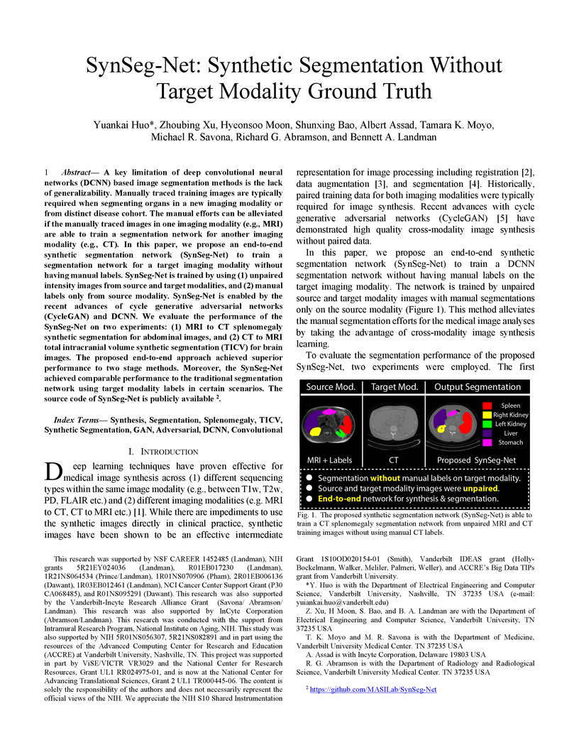
propose an end-to-end synthetic segmentation network (SynSeg-Net) to train a
segmentation network for a target imaging modality without having manual
labels. SynSeg-Net is trained by using (1) unpaired intensity images from
source and target modalities, and (2) manual labels only from source modality.
SynSeg-Net is enabled by the recent advances of cycle generative adversarial
networks (CycleGAN) and DCNN. We evaluate the performance of the SynSeg-Net on
two experiments: (1) MRI to CT splenomegaly synthetic segmentation for
abdominal images, and (2) CT to MRI total intracranial volume synthetic
segmentation (TICV) for brain images. The proposed end-to-end approach achieved
superior performance to two stage methods. Moreover, the SynSeg-Net achieved
comparable performance to the traditional segmentation network using target
modality labels in certain scenarios. The source code of SynSeg-Net is publicly
available (this https URL).
14 Mar 2024

Social science NLP tasks, such as emotion or humor detection, are required to capture the semantics along with the implicit pragmatics from text, often with limited amounts of training data. Instruction tuning has been shown to improve the many capabilities of large language models (LLMs) such as commonsense reasoning, reading comprehension, and computer programming. However, little is known about the effectiveness of instruction tuning on the social domain where implicit pragmatic cues are often needed to be captured. We explore the use of instruction tuning for social science NLP tasks and introduce Socialite-Llama -- an open-source, instruction-tuned Llama. On a suite of 20 social science tasks, Socialite-Llama improves upon the performance of Llama as well as matches or improves upon the performance of a state-of-the-art, multi-task finetuned model on a majority of them. Further, Socialite-Llama also leads to improvement on 5 out of 6 related social tasks as compared to Llama, suggesting instruction tuning can lead to generalized social understanding. All resources including our code, model and dataset can be found through this http URL.
25 Apr 2023

The lack of standardization is a prominent issue in magnetic resonance (MR) imaging. This often causes undesired contrast variations in the acquired images due to differences in hardware and acquisition parameters. In recent years, image synthesis-based MR harmonization with disentanglement has been proposed to compensate for the undesired contrast variations. Despite the success of existing methods, we argue that three major improvements can be made. First, most existing methods are built upon the assumption that multi-contrast MR images of the same subject share the same anatomy. This assumption is questionable, since different MR contrasts are specialized to highlight different anatomical features. Second, these methods often require a fixed set of MR contrasts for training (e.g., both T1-weighted and T2-weighted images), limiting their applicability. Lastly, existing methods are generally sensitive to imaging artifacts. In this paper, we present Harmonization with Attention-based Contrast, Anatomy, and Artifact Awareness (HACA3), a novel approach to address these three issues. HACA3 incorporates an anatomy fusion module that accounts for the inherent anatomical differences between MR contrasts. Furthermore, HACA3 is also robust to imaging artifacts and can be trained and applied to any set of MR contrasts. HACA3 is developed and evaluated on diverse MR datasets acquired from 21 sites with varying field strengths, scanner platforms, and acquisition protocols. Experiments show that HACA3 achieves state-of-the-art performance under multiple image quality metrics. We also demonstrate the applicability and versatility of HACA3 on downstream tasks including white matter lesion segmentation and longitudinal volumetric analyses.
30 Sep 2024
AI in Medical Imaging project aims to enhance the National Cancer Institute's
(NCI) Image Data Commons (IDC) by developing nnU-Net models and providing
AI-assisted segmentations for cancer radiology images. We created high-quality,
AI-annotated imaging datasets for 11 IDC collections. These datasets include
images from various modalities, such as computed tomography (CT) and magnetic
resonance imaging (MRI), covering the lungs, breast, brain, kidneys, prostate,
and liver. The nnU-Net models were trained using open-source datasets. A
portion of the AI-generated annotations was reviewed and corrected by
radiologists. Both the AI and radiologist annotations were encoded in
compliance with the the Digital Imaging and Communications in Medicine (DICOM)
standard, ensuring seamless integration into the IDC collections. All models,
images, and annotations are publicly accessible, facilitating further research
and development in cancer imaging. This work supports the advancement of
imaging tools and algorithms by providing comprehensive and accurate annotated
datasets.
31 Jul 2025
UCLAStanford UniversityShanghaiTech UniversityDeloitte Consulting LLPNational Tsing-Hua UniversityGE HealthCareUniversity of South FloridaGerman Cancer Research CenterCedars-Sinai Medical CenterVanderbilt University Medical CenterRowan UniversityHeidelberg University HospitalIcahn School of Medicine at Mount SinaiAmazon Web Services (AWS)Frederick National Laboratory for Cancer ResearchUniversity Medical Center FreiburgNational Cancer InstituteMoffitt Cancer CenterNational Institute of HealthNational Heart Lung and Blood InstituteSage BionetworksPixelMed PublishingUniversity of Arkansas for Medical SciencesInstitute of Cancer ResearchEssential Software Inc.Impact Business Information Solutions, IncEllumen, Inc.The de-identification (deID) of protected health information (PHI) and personally identifiable information (PII) is a fundamental requirement for sharing medical images, particularly through public repositories, to ensure compliance with patient privacy laws. In addition, preservation of non-PHI metadata to inform and enable downstream development of imaging artificial intelligence (AI) is an important consideration in biomedical research. The goal of MIDI-B was to provide a standardized platform for benchmarking of DICOM image deID tools based on a set of rules conformant to the HIPAA Safe Harbor regulation, the DICOM Attribute Confidentiality Profiles, and best practices in preservation of research-critical metadata, as defined by The Cancer Imaging Archive (TCIA). The challenge employed a large, diverse, multi-center, and multi-modality set of real de-identified radiology images with synthetic PHI/PII inserted.
The MIDI-B Challenge consisted of three phases: training, validation, and test. Eighty individuals registered for the challenge. In the training phase, we encouraged participants to tune their algorithms using their in-house or public data. The validation and test phases utilized the DICOM images containing synthetic identifiers (of 216 and 322 subjects, respectively). Ten teams successfully completed the test phase of the challenge. To measure success of a rule-based approach to image deID, scores were computed as the percentage of correct actions from the total number of required actions. The scores ranged from 97.91% to 99.93%. Participants employed a variety of open-source and proprietary tools with customized configurations, large language models, and optical character recognition (OCR). In this paper we provide a comprehensive report on the MIDI-B Challenge's design, implementation, results, and lessons learned.
29 Oct 2024

Estimated brain age from magnetic resonance image (MRI) and its deviation from chronological age can provide early insights into potential neurodegenerative diseases, supporting early detection and implementation of prevention strategies. Diffusion MRI (dMRI), a widely used modality for brain age estimation, presents an opportunity to build an earlier biomarker for neurodegenerative disease prediction because it captures subtle microstructural changes that precede more perceptible macrostructural changes. However, the coexistence of macro- and micro-structural information in dMRI raises the question of whether current dMRI-based brain age estimation models are leveraging the intended microstructural information or if they inadvertently rely on the macrostructural information. To develop a microstructure-specific brain age, we propose a method for brain age identification from dMRI that minimizes the model's use of macrostructural information by non-rigidly registering all images to a standard template. Imaging data from 13,398 participants across 12 datasets were used for the training and evaluation. We compare our brain age models, trained with and without macrostructural information minimized, with an architecturally similar T1-weighted (T1w) MRI-based brain age model and two state-of-the-art T1w MRI-based brain age models that primarily use macrostructural information. We observe difference between our dMRI-based brain age and T1w MRI-based brain age across stages of neurodegeneration, with dMRI-based brain age being older than T1w MRI-based brain age in participants transitioning from cognitively normal (CN) to mild cognitive impairment (MCI), but younger in participants already diagnosed with Alzheimer's disease (AD). Approximately 4 years before MCI diagnosis, dMRI-based brain age yields better performance than T1w MRI-based brain ages in predicting transition from CN to MCI.
31 Jul 2022
The majority of deep learning (DL) based deformable image registration methods use convolutional neural networks (CNNs) to estimate displacement fields from pairs of moving and fixed images. This, however, requires the convolutional kernels in the CNN to not only extract intensity features from the inputs but also understand image coordinate systems. We argue that the latter task is challenging for traditional CNNs, limiting their performance in registration tasks. To tackle this problem, we first introduce Coordinate Translator, a differentiable module that identifies matched features between the fixed and moving image and outputs their coordinate correspondences without the need for training. It unloads the burden of understanding image coordinate systems for CNNs, allowing them to focus on feature extraction. We then propose a novel deformable registration network, im2grid, that uses multiple Coordinate Translator's with the hierarchical features extracted from a CNN encoder and outputs a deformation field in a coarse-to-fine fashion. We compared im2grid with the state-of-the-art DL and non-DL methods for unsupervised 3D magnetic resonance image registration. Our experiments show that im2grid outperforms these methods both qualitatively and quantitatively.
05 Jun 2018
Whole brain segmentation on a structural magnetic resonance imaging (MRI) is essential in non-invasive investigation for neuroanatomy. Historically, multi-atlas segmentation (MAS) has been regarded as the de facto standard method for whole brain segmentation. Recently, deep neural network approaches have been applied to whole brain segmentation by learning random patches or 2D slices. Yet, few previous efforts have been made on detailed whole brain segmentation using 3D networks due to the following challenges: (1) fitting entire whole brain volume into 3D networks is restricted by the current GPU memory, and (2) the large number of targeting labels (e.g., > 100 labels) with limited number of training 3D volumes (e.g., < 50 scans). In this paper, we propose the spatially localized atlas network tiles (SLANT) method to distribute multiple independent 3D fully convolutional networks to cover overlapped sub-spaces in a standard atlas space. This strategy simplifies the whole brain learning task to localized sub-tasks, which was enabled by combing canonical registration and label fusion techniques with deep learning. To address the second challenge, auxiliary labels on 5111 initially unlabeled scans were created by MAS for pre-training. From empirical validation, the state-of-the-art MAS method achieved mean Dice value of 0.76, 0.71, and 0.68, while the proposed method achieved 0.78, 0.73, and 0.71 on three validation cohorts. Moreover, the computational time reduced from > 30 hours using MAS to ~15 minutes using the proposed method. The source code is available online this https URL
11 Mar 2019
Machine learning models are becoming commonplace in the domain of medical imaging, and with these methods comes an ever-increasing need for more data. However, to preserve patient anonymity it is frequently impractical or prohibited to transfer protected health information (PHI) between institutions. Additionally, due to the nature of some studies, there may not be a large public dataset available on which to train models. To address this conundrum, we analyze the efficacy of transferring the model itself in lieu of data between different sites. By doing so we accomplish two goals: 1) the model gains access to training on a larger dataset that it could not normally obtain and 2) the model better generalizes, having trained on data from separate locations. In this paper, we implement multi-site learning with disparate datasets from the National Institutes of Health (NIH) and Vanderbilt University Medical Center (VUMC) without compromising PHI. Three neural networks are trained to convergence on a computed tomography (CT) brain hematoma segmentation task: one only with NIH data,one only with VUMC data, and one multi-site model alternating between NIH and VUMC data. Resultant lesion masks with the multi-site model attain an average Dice similarity coefficient of 0.64 and the automatically segmented hematoma volumes correlate to those done manually with a Pearson correlation coefficient of 0.87,corresponding to an 8% and 5% improvement, respectively, over the single-site model counterparts.

12 Jan 2025
By 2030, the senior population aged 65 and older is expected to increase by over 50%, significantly raising the number of older drivers on the road. Drivers over 70 face higher crash death rates compared to those in their forties and fifties, underscoring the importance of developing more effective safety interventions for this demographic. Although the impact of aging on driving behavior has been studied, there is limited research on how these behaviors translate into real-world driving scenarios. This study addresses this need by leveraging Naturalistic Driving Data (NDD) to analyze driving performance measures - specifically, speed limit adherence on interstates and deceleration at stop intersections, both of which may be influenced by age-related declines. Using NDD, we developed Cumulative Distribution Functions (CDFs) to establish benchmarks for key driving behaviors among senior and young drivers. Our analysis, which included anomaly detection, benchmark comparisons, and accuracy evaluations, revealed significant differences in driving patterns primarily related to speed limit adherence at 75mph. While our approach shows promising potential for enhancing Advanced Driver Assistance Systems (ADAS) by providing tailored interventions based on age-specific adherence to speed limit driving patterns, we recognize the need for additional data to refine and validate metrics for other driving behaviors. By establishing precise benchmarks for various driving performance metrics, ADAS can effectively identify anomalies, such as abrupt deceleration, which may indicate impaired driving or other safety concerns. This study lays a strong foundation for future research aimed at improving safety interventions through detailed driving behavior analysis.
31 Jan 2025
Defacing is often applied to head magnetic resonance image (MRI) datasets prior to public release to address privacy concerns. The alteration of facial and nearby voxels has provoked discussions about the true capability of these techniques to ensure privacy as well as their impact on downstream tasks. With advancements in deep generative models, the extent to which defacing can protect privacy is uncertain. Additionally, while the altered voxels are known to contain valuable anatomical information, their potential to support research beyond the anatomical regions directly affected by defacing remains uncertain. To evaluate these considerations, we develop a refacing pipeline that recovers faces in defaced head MRIs using cascaded diffusion probabilistic models (DPMs). The DPMs are trained on images from 180 subjects and tested on images from 484 unseen subjects, 469 of whom are from a different dataset. To assess whether the altered voxels in defacing contain universally useful information, we also predict computed tomography (CT)-derived skeletal muscle radiodensity from facial voxels in both defaced and original MRIs. The results show that DPMs can generate high-fidelity faces that resemble the original faces from defaced images, with surface distances to the original faces significantly smaller than those of a population average face (p < 0.05). This performance also generalizes well to previously unseen datasets. For skeletal muscle radiodensity predictions, using defaced images results in significantly weaker Spearman's rank correlation coefficients compared to using original images (p < 10-4). For shin muscle, the correlation is statistically significant (p < 0.05) when using original images but not statistically significant (p > 0.05) when any defacing method is applied, suggesting that defacing might not only fail to protect privacy but also eliminate valuable information.
16 Nov 2017

Background. Wearable accelerometry devices allow collection of high-density
activity data in large epidemiological studies both in-the-lab as well as
in-the-wild (free-living). Such data can be used to detect and identify periods
of sustained harmonic walking. This report aims to establish whether the micro-
and macro-features of walking identified in the laboratory and free-living
environments are associated with measures of physical function, mobility,
fatigability, and fitness.
Methods. Fifty-one older adults (median age 77.5) enrolled in the
Developmental Epidemiologic Cohort Study in Pittsburgh, Pennsylvania were
included in the analyses. The study included an in-the-lab component as well as
7 days of monitoring in-the-wild. Participants were equipped with hip-worn
Actigraph GT3X+ activity monitors, which collect high-density raw accelerometry
data. We applied a walking identification algorithm to the data and defined
features of walking, such as participant-specific walking acceleration and
cadence. The association between these walking features and physical function,
mobility, fatigability, and fitness was quantified using linear regression
analysis.
Results. Micro-scale features of walking (acceleration and cadence) estimated
from in-the-lab and in-the-wild data were associated with measures of physical
function, mobility, fatigability, and fitness. In-the-lab median walking
acceleration was strongly inversely associated with physical function,
mobility, fatigability and fitness. Additionally, in-the-wild daily walking
time was inversely associated with usual- and fast-paced 400m walking time.
Conclusions. The proposed accelerometry-derived walking features are
significantly associated with measures of physical function, mobility,
fatigability, and fitness, which provides evidence of convergent validity.
01 Jun 2016
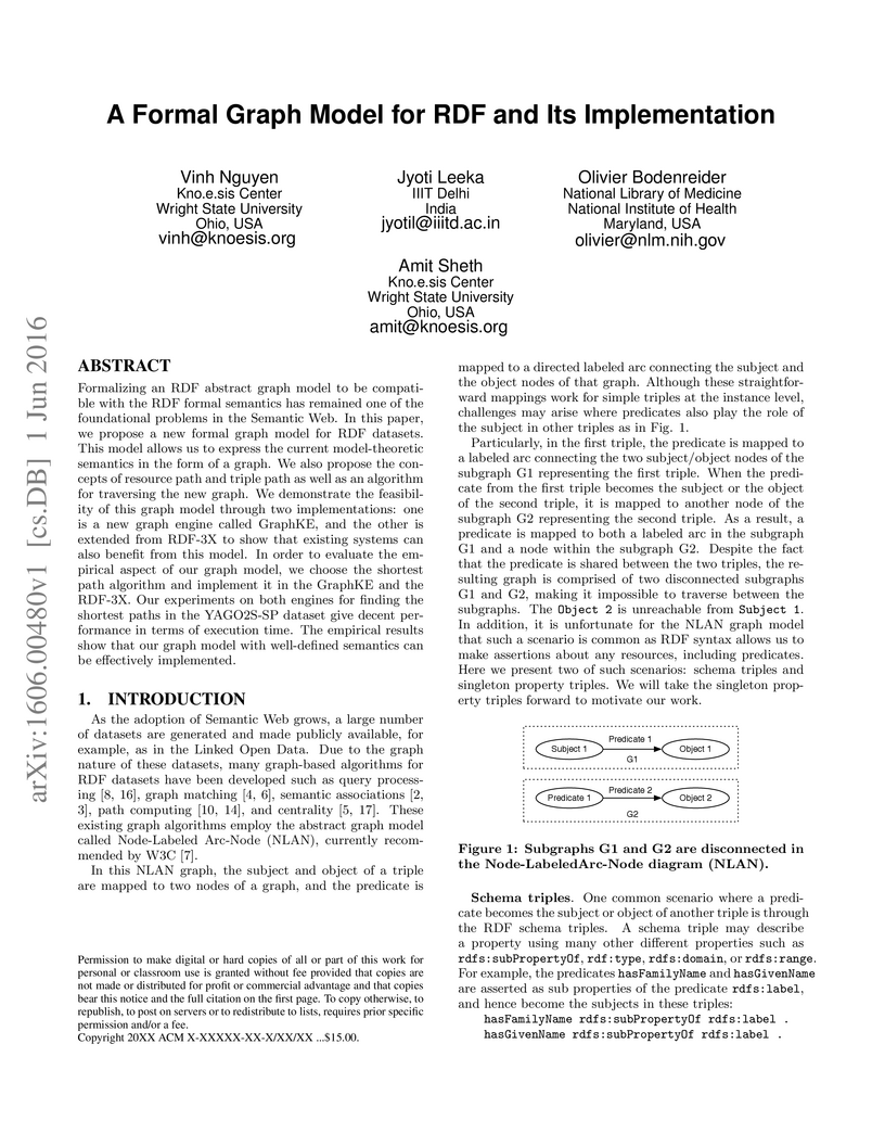
Formalizing an RDF abstract graph model to be compatible with the RDF formal
semantics has remained one of the foundational problems in the Semantic Web. In
this paper, we propose a new formal graph model for RDF datasets. This model
allows us to express the current model-theoretic semantics in the form of a
graph. We also propose the concepts of resource path and triple path as well as
an algorithm for traversing the new graph. We demonstrate the feasibility of
this graph model through two implementations: one is a new graph engine called
GraphKE, and the other is extended from RDF-3X to show that existing systems
can also benefit from this model. In order to evaluate the empirical aspect of
our graph model, we choose the shortest path algorithm and implement it in the
GraphKE and the RDF-3X. Our experiments on both engines for finding the
shortest paths in the YAGO2S-SP dataset give decent performance in terms of
execution time. The empirical results show that our graph model with
well-defined semantics can be effectively implemented.
27 Jul 2018
With the increasing popularity of PET-MR scanners in clinical applications,
synthesis of CT images from MR has been an important research topic. Accurate
PET image reconstruction requires attenuation correction, which is based on the
electron density of tissues and can be obtained from CT images. While CT
measures electron density information for x-ray photons, MR images convey
information about the magnetic properties of tissues. Therefore, with the
advent of PET-MR systems, the attenuation coefficients need to be indirectly
estimated from MR images. In this paper, we propose a fully convolutional
neural network (CNN) based method to synthesize head CT from ultra-short
echo-time (UTE) dual-echo MR images. Unlike traditional -w images which do
not have any bone signal, UTE images show some signal for bone, which makes it
a good candidate for MR to CT synthesis. A notable advantage of our approach is
that accurate results were achieved with a small training data set. Using an
atlas of a single CT and dual-echo UTE pair, we train a deep neural network
model to learn the transform of MR intensities to CT using patches. We compared
our CNN based model with a state-of-the-art registration based as well as a
Bayesian model based CT synthesis method, and showed that the proposed CNN
model outperforms both of them. We also compared the proposed model when only
-w images are available instead of UTE, and show that UTE images produce
better synthesis than using just -w images.
03 Dec 2024

Diffusion-weighted magnetic resonance imaging (DW-MRI) is a critical imaging
method for capturing and modeling tissue microarchitecture at a millimeter
scale. A common practice to model the measured DW-MRI signal is via fiber
orientation distribution function (fODF). This function is the essential first
step for the downstream tractography and connectivity analyses. With recent
advantages in data sharing, large-scale multi-site DW-MRI datasets are being
made available for multi-site studies. However, measurement variabilities
(e.g., inter- and intra-site variability, hardware performance, and sequence
design) are inevitable during the acquisition of DW-MRI. Most existing
model-based methods (e.g., constrained spherical deconvolution (CSD)) and
learning based methods (e.g., deep learning (DL)) do not explicitly consider
such variabilities in fODF modeling, which consequently leads to inferior
performance on multi-site and/or longitudinal diffusion studies. In this paper,
we propose a novel data-driven deep constrained spherical deconvolution method
to explicitly constrain the scan-rescan variabilities for a more reproducible
and robust estimation of brain microstructure from repeated DW-MRI scans.
Specifically, the proposed method introduces a new 3D volumetric
scanner-invariant regularization scheme during the fODF estimation. We study
the Human Connectome Project (HCP) young adults test-retest group as well as
the MASiVar dataset (with inter- and intra-site scan/rescan data). The
Baltimore Longitudinal Study of Aging (BLSA) dataset is employed for external
validation. From the experimental results, the proposed data-driven framework
outperforms the existing benchmarks in repeated fODF estimation. The proposed
method is assessing the downstream connectivity analysis and shows increased
performance in distinguishing subjects with different biomarkers.
25 Jan 2023
CSIROUniversity of Southern CaliforniaThe University of MelbourneUniversity of PennsylvaniaWashington University in St. LouisJohns Hopkins University School of MedicineUniversity of LausanneUniversity of Wisconsin School of Medicine and Public HealthNational Institute on AgingNational Institutes of HealthLausanne University HospitalUniversity of Texas San Antonio Health Science Center
University of Southern CaliforniaThe University of MelbourneUniversity of PennsylvaniaWashington University in St. LouisJohns Hopkins University School of MedicineUniversity of LausanneUniversity of Wisconsin School of Medicine and Public HealthNational Institute on AgingNational Institutes of HealthLausanne University HospitalUniversity of Texas San Antonio Health Science CenterDisease heterogeneity has been a critical challenge for precision diagnosis and treatment, especially in neurologic and neuropsychiatric diseases. Many diseases can display multiple distinct brain phenotypes across individuals, potentially reflecting disease subtypes that can be captured using MRI and machine learning methods. However, biological interpretability and treatment relevance are limited if the derived subtypes are not associated with genetic drivers or susceptibility factors. Herein, we describe Gene-SGAN - a multi-view, weakly-supervised deep clustering method - which dissects disease heterogeneity by jointly considering phenotypic and genetic data, thereby conferring genetic correlations to the disease subtypes and associated endophenotypic signatures. We first validate the generalizability, interpretability, and robustness of Gene-SGAN in semi-synthetic experiments. We then demonstrate its application to real multi-site datasets from 28,858 individuals, deriving subtypes of Alzheimer's disease and brain endophenotypes associated with hypertension, from MRI and SNP data. Derived brain phenotypes displayed significant differences in neuroanatomical patterns, genetic determinants, biological and clinical biomarkers, indicating potentially distinct underlying neuropathologic processes, genetic drivers, and susceptibility factors. Overall, Gene-SGAN is broadly applicable to disease subtyping and endophenotype discovery, and is herein tested on disease-related, genetically-driven neuroimaging phenotypes.
28 Sep 2022
Metabolic health is increasingly implicated as a risk factor across
conditions from cardiology to neurology, and efficiency assessment of body
composition is critical to quantitatively characterizing these relationships.
2D low dose single slice computed tomography (CT) provides a high resolution,
quantitative tissue map, albeit with a limited field of view. Although numerous
potential analyses have been proposed in quantifying image context, there has
been no comprehensive study for low-dose single slice CT longitudinal
variability with automated segmentation. We studied a total of 1816 slices from
1469 subjects of Baltimore Longitudinal Study on Aging (BLSA) abdominal dataset
using supervised deep learning-based segmentation and unsupervised clustering
method. 300 out of 1469 subjects that have two year gap in their first two
scans were pick out to evaluate longitudinal variability with measurements
including intraclass correlation coefficient (ICC) and coefficient of variation
(CV) in terms of tissues/organs size and mean intensity. We showed that our
segmentation methods are stable in longitudinal settings with Dice ranged from
0.821 to 0.962 for thirteen target abdominal tissues structures. We observed
high variability in most organ with ICC<0.5, low variability in the area of
muscle, abdominal wall, fat and body mask with average ICC>0.8. We found that
the variability in organ is highly related to the cross-sectional position of
the 2D slice. Our efforts pave quantitative exploration and quality control to
reduce uncertainties in longitudinal analysis.
There are no more papers matching your filters at the moment.
