07 Apr 2025


Researchers from Emory University and partner institutions develop Collab-RAG, a framework that enables efficient collaboration between small and large language models for complex question answering, achieving 1.8-14.2% performance improvements across five multi-hop QA datasets through automated query decomposition and iterative preference optimization.
27 Jun 2023
The LViT model, developed by researchers from Xiamen University, UIUC, UT Southwestern Medical Center, and Alibaba's DAMO Academy, introduces a text-augmented Vision Transformer for medical image segmentation. This model effectively integrates medical text reports to improve segmentation accuracy and data efficiency, achieving superior performance on CT and X-ray datasets while significantly reducing reliance on extensive pixel-level annotations.
27 Aug 2025
This survey critically evaluates the progress in deep learning-based medical image segmentation from 2015 to 2024, concluding that while performance has improved significantly, the problem remains unsolved, especially concerning clinical viability and generalization. It identifies persistent challenges across various segmentation paradigms and outlines future research directions, including the development of intelligent segmentation agents and the need for new evaluation metrics that quantify human-in-the-loop efficiency.
21 Aug 2025


Three-Dimensional Gaussian Splatting (3DGS) has shown substantial promise in the field of computer vision, but remains unexplored in the field of magnetic resonance imaging (MRI). This study explores its potential for the reconstruction of isotropic resolution 3D MRI from undersampled k-space data. We introduce a novel framework termed 3D Gaussian MRI (3DGSMR), which employs 3D Gaussian distributions as an explicit representation for MR volumes. Experimental evaluations indicate that this method can effectively reconstruct voxelized MR images, achieving a quality on par with that of well-established 3D MRI reconstruction techniques found in the literature. Notably, the 3DGSMR scheme operates under a self-supervised framework, obviating the need for extensive training datasets or prior model training. This approach introduces significant innovations to the domain, notably the adaptation of 3DGS to MRI reconstruction and the novel application of the existing 3DGS methodology to decompose MR signals, which are presented in a complex-valued format.
23 Nov 2024
Diffusion models have demonstrated significant potential in producing
high-quality images in medical image translation to aid disease diagnosis,
localization, and treatment. Nevertheless, current diffusion models have
limited success in achieving faithful image translations that can accurately
preserve the anatomical structures of medical images, especially for unpaired
datasets. The preservation of structural and anatomical details is essential to
reliable medical diagnosis and treatment planning, as structural mismatches can
lead to disease misidentification and treatment errors. In this study, we
introduce the Frequency Decoupled Diffusion Model (FDDM) for MR-to-CT
conversion. FDDM first obtains the anatomical information of the CT image from
the MR image through an initial conversion module. This anatomical information
then guides a subsequent diffusion model to generate high-quality CT images.
Our diffusion model uses a dual-path reverse diffusion process for
low-frequency and high-frequency information, achieving a better balance
between image quality and anatomical accuracy. We extensively evaluated FDDM
using public datasets for brain MR-to-CT and pelvis MR-to-CT translations,
demonstrating its superior performance to other GAN-based, VAE-based, and
diffusion-based models. The evaluation metrics included Frechet Inception
Distance (FID), Peak Signal-to-Noise Ratio (PSNR), and Structural Similarity
Index Measure (SSIM). FDDM achieved the best scores on all metrics for both
datasets, particularly excelling in FID, with scores of 25.9 for brain data and
29.2 for pelvis data, significantly outperforming other methods. These results
demonstrate that FDDM can generate high-quality target domain images while
maintaining the accuracy of translated anatomical structures.
01 Aug 2025
This paper offers a comprehensive review of Video Object Segmentation and Tracking (VOST) methods, particularly focusing on the impact and integration of foundation models like the Segment Anything Model (SAM) and its successor, SAM2. It systematically analyzes how these models address challenges in memory management, feature learning, and trajectory estimation across different temporal stages, outlining key future research directions for building more robust and clinically applicable VOST systems, especially in medical imaging.
09 Oct 2024
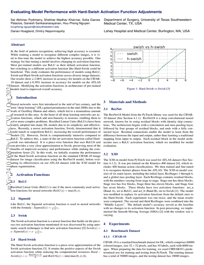
In the field of pattern recognition, achieving high accuracy is essential. While training a model to recognize different complex images, it is vital to fine-tune the model to achieve the highest accuracy possible. One strategy for fine-tuning a model involves changing its activation function. Most pre-trained models use ReLU as their default activation function, but switching to a different activation function like Hard-Swish could be beneficial. This study evaluates the performance of models using ReLU, Swish and Hard-Swish activation functions across diverse image datasets. Our results show a 2.06% increase in accuracy for models on the CIFAR-10 dataset and a 0.30% increase in accuracy for models on the ATLAS dataset. Modifying the activation functions in architecture of pre-trained models lead to improved overall accuracy.
15 May 2024

Automatic segmentation of medical images is crucial in modern clinical
workflows. The Segment Anything Model (SAM) has emerged as a versatile tool for
image segmentation without specific domain training, but it requires human
prompts and may have limitations in specific domains. Traditional models like
nnUNet perform automatic segmentation during inference and are effective in
specific domains but need extensive domain-specific training. To combine the
strengths of foundational and domain-specific models, we propose nnSAM,
integrating SAM's robust feature extraction with nnUNet's automatic
configuration to enhance segmentation accuracy on small datasets. Our nnSAM
model optimizes two main approaches: leveraging SAM's feature extraction and
nnUNet's domain-specific adaptation, and incorporating a boundary shape
supervision loss function based on level set functions and curvature
calculations to learn anatomical shape priors from limited data. We evaluated
nnSAM on four segmentation tasks: brain white matter, liver, lung, and heart
segmentation. Our method outperformed others, achieving the highest DICE score
of 82.77% and the lowest ASD of 1.14 mm in brain white matter segmentation with
20 training samples, compared to nnUNet's DICE score of 79.25% and ASD of 1.36
mm. A sample size study highlighted nnSAM's advantage with fewer training
samples. Our results demonstrate significant improvements in segmentation
performance with nnSAM, showcasing its potential for small-sample learning in
medical image segmentation.
08 May 2025
This study presents an unsupervised, motion-resolved reconstruction framework
for high-resolution, free-breathing pulmonary magnetic resonance imaging (MRI),
utilizing a three-dimensional Gaussian representation (3DGS). The proposed
method leverages 3DGS to address the challenges of motion-resolved 3D isotropic
pulmonary MRI reconstruction by enabling data smoothing between voxels for
continuous spatial representation. Pulmonary MRI data acquisition is performed
using a golden-angle radial sampling trajectory, with respiratory motion
signals extracted from the center of k-space in each radial spoke. Based on the
estimated motion signal, the k-space data is sorted into multiple respiratory
phases. A 3DGS framework is then applied to reconstruct a reference image
volume from the first motion state. Subsequently, a patient-specific
convolutional neural network is trained to estimate the deformation vector
fields (DVFs), which are used to generate the remaining motion states through
spatial transformation of the reference volume. The proposed reconstruction
pipeline is evaluated on six datasets from six subjects and bench-marked
against three state-of-the-art reconstruction methods. The experimental
findings demonstrate that the proposed reconstruction framework effectively
reconstructs high-resolution, motion-resolved pulmonary MR images. Compared
with existing approaches, it achieves superior image quality, reflected by
higher signal-to-noise ratio and contrast-to-noise ratio. The proposed
unsupervised 3DGS-based reconstruction method enables accurate motion-resolved
pulmonary MRI with isotropic spatial resolution. Its superior performance in
image quality metrics over state-of-the-art methods highlights its potential as
a robust solution for clinical pulmonary MR imaging.
17 Feb 2025
Inverse generation problems, such as denoising without ground truth
observations, is a critical challenge in many scientific inquiries and
real-world applications. While recent advances in generative models like
diffusion models, conditional flow matching, and consistency models achieved
impressive results by casting generation as denoising problems, they cannot be
directly used for inverse generation without access to clean data. Here we
introduce Inverse Flow (IF), a novel framework that enables using these
generative models for inverse generation problems including denoising without
ground truth. Inverse Flow can be flexibly applied to nearly any continuous
noise distribution and allows complex dependencies. We propose two algorithms
for learning Inverse Flows, Inverse Flow Matching (IFM) and Inverse Consistency
Model (ICM). Notably, to derive the computationally efficient, simulation-free
inverse consistency model objective, we generalized consistency training to any
forward diffusion processes or conditional flows, which have applications
beyond denoising. We demonstrate the effectiveness of IF on synthetic and real
datasets, outperforming prior approaches while enabling noise distributions
that previous methods cannot support. Finally, we showcase applications of our
techniques to fluorescence microscopy and single-cell genomics data,
highlighting IF's utility in scientific problems. Overall, this work expands
the applications of powerful generative models to inversion generation
problems.
20 Aug 2025
Tibetan is a low-resource language with minimal parallel speech corpora spanning its three major dialects-Ü-Tsang, Amdo, and Kham-limiting progress in speech modeling. To address this issue, we propose FMSD-TTS, a few-shot, multi-speaker, multi-dialect text-to-speech framework that synthesizes parallel dialectal speech from limited reference audio and explicit dialect labels. Our method features a novel speaker-dialect fusion module and a Dialect-Specialized Dynamic Routing Network (DSDR-Net) to capture fine-grained acoustic and linguistic variations across dialects while preserving speaker identity. Extensive objective and subjective evaluations demonstrate that FMSD-TTS significantly outperforms baselines in both dialectal expressiveness and speaker similarity. We further validate the quality and utility of the synthesized speech through a challenging speech-to-speech dialect conversion task. Our contributions include: (1) a novel few-shot TTS system tailored for Tibetan multi-dialect speech synthesis, (2) the public release of a large-scale synthetic Tibetan speech corpus generated by FMSD-TTS, and (3) an open-source evaluation toolkit for standardized assessment of speaker similarity, dialect consistency, and audio quality.
17 Sep 2025
AI-based in silico methods have improved protein structure prediction but often struggle with large protein complexes (PCs) involving multiple interacting proteins due to missing 3D spatial cues. Experimental techniques like Cryo-EM are accurate but costly and time-consuming. We present ProFusion, a hybrid framework that integrates a deep learning model with Atomic Force Microscopy (AFM), which provides high-resolution height maps from random orientations, naturally yielding multi-view data for 3D reconstruction. However, generating a large-scale AFM imaging data set sufficient to train deep learning models is impractical. Therefore, we developed a virtual AFM framework that simulates the imaging process and generated a dataset of ~542,000 proteins with multi-view synthetic AFM images. We train a conditional diffusion model to synthesize novel views from unposed inputs and an instance-specific Neural Radiance Field (NeRF) model to reconstruct 3D structures. Our reconstructed 3D protein structures achieve an average Chamfer Distance within the AFM imaging resolution, reflecting high structural fidelity. Our method is extensively validated on experimental AFM images of various PCs, demonstrating strong potential for accurate, cost-effective protein complex structure prediction and rapid iterative validation using AFM experiments.
25 Jul 2025
Purpose: Accurate tumor segmentation is vital for adaptive radiation therapy (ART) but remains time-consuming and user-dependent. Segment Anything Model 2 (SAM2) shows promise for prompt-based segmentation but struggles with tumor accuracy. We propose prior knowledge-based augmentation strategies to enhance SAM2 for ART.
Methods: Two strategies were introduced to improve SAM2: (1) using prior MR images and annotations as contextual inputs, and (2) improving prompt robustness via random bounding box expansion and mask erosion/dilation. The resulting model, SAM2-Aug, was fine-tuned and tested on the One-Seq-Liver dataset (115 MRIs from 31 liver cancer patients), and evaluated without retraining on Mix-Seq-Abdomen (88 MRIs, 28 patients) and Mix-Seq-Brain (86 MRIs, 37 patients).
Results: SAM2-Aug outperformed convolutional, transformer-based, and prompt-driven models across all datasets, achieving Dice scores of 0.86(liver), 0.89(abdomen), and 0.90(brain). It demonstrated strong generalization across tumor types and imaging sequences, with improved performance in boundary-sensitive metrics.
Conclusions: Incorporating prior images and enhancing prompt diversity significantly boosts segmentation accuracy and generalizability. SAM2-Aug offers a robust, efficient solution for tumor segmentation in ART. Code and models will be released at this https URL.
25 Nov 2025

Dynamic positron emission tomography (PET) imaging combined with radiotracer kinetic modeling is a powerful technique for visualizing biological processes in the brain, offering valuable insights into brain functions and neurological disorders such as Alzheimer's and Parkinson's diseases. Accurate kinetic modeling relies heavily on the use of a metabolite-corrected arterial input function (AIF), which typically requires invasive and labor-intensive arterial blood sampling. While alternative non-invasive approaches have been proposed, they often compromise accuracy or still necessitate at least one invasive blood sampling. In this study, we present the deep learning-derived arterial input function (DLIF), a deep learning framework capable of estimating a metabolite-corrected AIF directly from dynamic PET image sequences without any blood sampling. We validated DLIF using existing dynamic PET patient data. We compared DLIF and resulting parametric maps against ground truth measurements. Our evaluation shows that DLIF achieves accurate and robust AIF estimation. By leveraging deep learning's ability to capture complex temporal dynamics and incorporating prior knowledge of typical AIF shapes through basis functions, DLIF provides a rapid, accurate, and entirely non-invasive alternative to traditional AIF measurement methods.
23 Aug 2022
 Imperial College LondonUniversity of Bern
 University of OxfordNanjing University of Science and Technology
 McGill University
 University of PennsylvaniaMassachusetts General HospitalUppsala UniversityUniversity of AucklandUniversity of Texas at DallasOld Dominion UniversityINSERMIndian Statistical InstituteUniversity of CalcuttaUniversidad de Los AndesUniversitat Polit`ecnica de CatalunyaUniversity of Texas Southwestern Medical CenterUme ̊a UniversityThe University of Pittsburgh Medical CenterUniversit
e de Tours
University of PennsylvaniaMassachusetts General HospitalUppsala UniversityUniversity of AucklandUniversity of Texas at DallasOld Dominion UniversityINSERMIndian Statistical InstituteUniversity of CalcuttaUniversidad de Los AndesUniversitat Polit`ecnica de CatalunyaUniversity of Texas Southwestern Medical CenterUme ̊a UniversityThe University of Pittsburgh Medical CenterUniversit
e de ToursDeep learning (DL) models have provided state-of-the-art performance in
various medical imaging benchmarking challenges, including the Brain Tumor
Segmentation (BraTS) challenges. However, the task of focal pathology
multi-compartment segmentation (e.g., tumor and lesion sub-regions) is
particularly challenging, and potential errors hinder translating DL models
into clinical workflows. Quantifying the reliability of DL model predictions in
the form of uncertainties could enable clinical review of the most uncertain
regions, thereby building trust and paving the way toward clinical translation.
Several uncertainty estimation methods have recently been introduced for DL
medical image segmentation tasks. Developing scores to evaluate and compare the
performance of uncertainty measures will assist the end-user in making more
informed decisions. In this study, we explore and evaluate a score developed
during the BraTS 2019 and BraTS 2020 task on uncertainty quantification
(QU-BraTS) and designed to assess and rank uncertainty estimates for brain
tumor multi-compartment segmentation. This score (1) rewards uncertainty
estimates that produce high confidence in correct assertions and those that
assign low confidence levels at incorrect assertions, and (2) penalizes
uncertainty measures that lead to a higher percentage of under-confident
correct assertions. We further benchmark the segmentation uncertainties
generated by 14 independent participating teams of QU-BraTS 2020, all of which
also participated in the main BraTS segmentation task. Overall, our findings
confirm the importance and complementary value that uncertainty estimates
provide to segmentation algorithms, highlighting the need for uncertainty
quantification in medical image analyses. Finally, in favor of transparency and
reproducibility, our evaluation code is made publicly available at:
this https URL

05 Oct 2016
Neural networks are known to be effective function approximators. Recently,
deep neural networks have proven to be very effective in pattern recognition,
classification tasks and human-level control to model highly nonlinear
realworld systems. This paper investigates the effectiveness of deep neural
networks in the modeling of dynamical systems with complex behavior. Three deep
neural network structures are trained on sequential data, and we investigate
the effectiveness of these networks in modeling associated characteristics of
the underlying dynamical systems. We carry out similar evaluations on select
publicly available system identification datasets. We demonstrate that deep
neural networks are effective model estimators from input-output data
18 Jul 2025
This study introduces a LLMs powered multiagent ensemble method to address challenges in hallucination and data labeling, particularly in large-scale EHR datasets. Manual labeling of such datasets requires domain expertise and is labor-intensive, time-consuming, expensive, and error-prone. To overcome this bottleneck, we developed an ensemble LLMs method and demonstrated its effectiveness in two real-world tasks: (1) labeling a large-scale unlabeled ECG dataset in MIMIC-IV; (2) identifying social determinants of health (SDOH) from the clinical notes of EHR. Trading off benefits and cost, we selected a pool of diverse open source LLMs with satisfactory performance. We treat each LLM's prediction as a vote and apply a mechanism of majority voting with minimal winning threshold for ensemble. We implemented an ensemble LLMs application for EHR data labeling tasks. By using the ensemble LLMs and natural language processing, we labeled MIMIC-IV ECG dataset of 623,566 ECG reports with an estimated accuracy of 98.2%. We applied the ensemble LLMs method to identify SDOH from social history sections of 1,405 EHR clinical notes, also achieving competitive performance. Our experiments show that the ensemble LLMs can outperform individual LLM even the best commercial one, and the method reduces hallucination errors. From the research, we found that (1) the ensemble LLMs method significantly reduces the time and effort required for labeling large-scale EHR data, automating the process with high accuracy and quality; (2) the method generalizes well to other text data labeling tasks, as shown by its application to SDOH identification; (3) the ensemble of a group of diverse LLMs can outperform or match the performance of the best individual LLM; and (4) the ensemble method substantially reduces hallucination errors. This approach provides a scalable and efficient solution to data-labeling challenges.
24 Jul 2025
Time-resolved CBCT imaging, which reconstructs a dynamic sequence of CBCTs reflecting intra-scan motion (one CBCT per x-ray projection without phase sorting or binning), is highly desired for regular and irregular motion characterization, patient setup, and motion-adapted radiotherapy. Representing patient anatomy and associated motion fields as 3D Gaussians, we developed a Gaussian representation-based framework (PMF-STGR) for fast and accurate dynamic CBCT reconstruction. PMF-STGR comprises three major components: a dense set of 3D Gaussians to reconstruct a reference-frame CBCT for the dynamic sequence; another 3D Gaussian set to capture three-level, coarse-to-fine motion-basis-components (MBCs) to model the intra-scan motion; and a CNN-based motion encoder to solve projection-specific temporal coefficients for the MBCs. Scaled by the temporal coefficients, the learned MBCs will combine into deformation vector fields to deform the reference CBCT into projection-specific, time-resolved CBCTs to capture the dynamic motion. Due to the strong representation power of 3D Gaussians, PMF-STGR can reconstruct dynamic CBCTs in a 'one-shot' training fashion from a standard 3D CBCT scan, without using any prior anatomical or motion model. We evaluated PMF-STGR using XCAT phantom simulations and real patient scans. Metrics including the image relative error, structural-similarity-index-measure, tumor center-of-mass-error, and landmark localization error were used to evaluate the accuracy of solved dynamic CBCTs and motion. PMF-STGR shows clear advantages over a state-of-the-art, INR-based approach, PMF-STINR. Compared with PMF-STINR, PMF-STGR reduces reconstruction time by 50% while reconstructing less blurred images with better motion accuracy. With improved efficiency and accuracy, PMF-STGR enhances the applicability of dynamic CBCT imaging for potential clinical translation.
15 Oct 2018
Deep learning has started to revolutionize several different industries, and
the applications of these methods in medicine are now becoming more
commonplace. This study focuses on investigating the feasibility of tracking
patients and clinical staff wearing Bluetooth Low Energy (BLE) tags in a
radiation oncology clinic using artificial neural networks (ANNs) and
convolutional neural networks (CNNs). The performance of these networks was
compared to relative received signal strength indicator (RSSI) thresholding and
triangulation. By utilizing temporal information, a combined CNN+ANN network
was capable of correctly identifying the location of the BLE tag with an
accuracy of 99.9%. It outperformed a CNN model (accuracy = 94%), a thresholding
model employing majority voting (accuracy = 95%), and a triangulation
classifier utilizing majority voting (accuracy = 95%). Future studies will seek
to deploy this affordable real time location system in hospitals to improve
clinical workflow, efficiency, and patient safety.
10 Dec 2024
Purpose: A reliable cancer prognosis model for clear cell renal cell carcinoma (ccRCC) can enhance personalized treatment. We developed a multi-modal ensemble model (MMEM) that integrates pretreatment clinical data, multi-omics data, and histopathology whole slide image (WSI) data to predict overall survival (OS) and disease-free survival (DFS) for ccRCC patients. Methods: We analyzed 226 patients from The Cancer Genome Atlas Kidney Renal Clear Cell Carcinoma (TCGA-KIRC) dataset, which includes OS, DFS follow-up data, and five data modalities: clinical data, WSIs, and three multi-omics datasets (mRNA, miRNA, and DNA methylation). Separate survival models were built for OS and DFS. Cox-proportional hazards (CPH) model with forward feature selection is used for clinical and multi-omics data. Features from WSIs were extracted using ResNet and three general-purpose foundation models. A deep learning-based CPH model predicted survival using encoded WSI features. Risk scores from all models were combined based on training performance. Results: Performance was assessed using concordance index (C-index) and AUROC. The clinical feature-based CPH model received the highest weight for both OS and DFS tasks. Among WSI-based models, the general-purpose foundation model (UNI) achieved the best performance. The final MMEM model surpassed single-modality models, achieving C-indices of 0.820 (OS) and 0.833 (DFS), and AUROC values of 0.831 (3-year patient death) and 0.862 (cancer recurrence). Using predicted risk medians to stratify high- and low-risk groups, log-rank tests showed improved performance in both OS and DFS compared to single-modality models. Conclusion: MMEM is the first multi-modal model for ccRCC patients, integrating five data modalities. It outperformed single-modality models in prognostic ability and has the potential to assist in ccRCC patient management if independently validated.
There are no more papers matching your filters at the moment.
